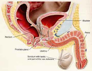
Nanda - Nursing Care Plan

Nursing Care Plan for Bone Cancer

Tumor is the growth of new cells, abnormal, progressive, where the cells never become mature. The incidence of bone tumors when compared with other types of tumors are small, ie less than 1% of all tumors of human body. Malignant tumor, when tumor capable of spreading to other places (able to metastasize) and benign tumors say, if it is not able to metastasize. Lungs, is the organ most frequently seized by child spread of malignant tumors.

There are many different types of cancer. Cancers are usually named based on the type of cell that is affected. For example, lung cancer is caused by cells that are beyond the control of the shape of the lung, and breast cancer by cells that form the breast. A tumor is a collection of abnormal cells which accumulate together. However, not all tumors are cancerous. A tumor can be benign (not cancerous) or malignant (cancerous).

Benign tumors are usually less dangerous and not able to spread to other parts of the body. Malignant tumors are generally more serious and can spread to other areas in the body. The ability of cancer cells to leave their original location and moved to another location in the body is called metastasis. Metastasis can occur with cancer cells enter the blood stream or lymphatic system body to walk to other places in the body.

When cancer cells metastasize to other parts of the body, they are still called by the type of origin of the abnormal cells. For example, if a group of cells into diseased breast cancer and metastasizes to the bones, it is called metastatic breast cancer. Many different types of cancer are able to metastasize to the bones.

The types of the most common cancers that spread to the bones are lung, breast, prostate, thyroid, and kidney. Most of the time, when people have cancer in their bones, it is caused by cancer that has spread from elsewhere in the body to the bones. It is less common to have an original bone cancer, a cancer that arises from cells that make bone. It is important to determine whether the cancer is in the bone from elsewhere or from a cancer of the bone cells. The treatments for cancers that have metastasized to the bone based on the initial type of cancer.

Causes

Bone cancer is caused by a problem with the cells that form bone. More than 2,000 people are diagnosed in the U.S. each year with a bone tumor. Bone tumors occur most commonly in children and adolescents and are less common in the older adults. Cancer involving the bone in adults older are most commonly the result of metastatic spread from another tumor.

Signs and Symptoms

The most common symptom of bone tumors is pain. In most cases, the symptoms become gradually more severe with time. At first, the pain may only be present at night or with activity. Depending on the growth of tumor, those affected may have symptoms for weeks, months, or years before seeking medical advice. In some cases, a mass or lump may be felt in the bone or in the tissues surrounding the bone. Tumors in the leg, causing the patient to walk lame, whereas tumors in the arm cause pain when the arm is used to lift some object. Swelling of the tumor may feel warm and slightly flushed.

Classification

Based on the level of malignancy, there are 3 levels of malignant tumor stages, namely:
  1. Stage I, when a low degree of malignancy.
  2. Stage II, meaning the tumor has a high degree of malignancy.
  3. Stage III, which means the tumor has spread.
Diagnostic tests
  1. Physical examination
  2. DPL
  3. X-Rays
  4. Ct-Scan
  5. MRI
  6. biopsy
  7. bone scan

Nursing Diagnosis for Bone Cancer

1. Anxiety related to change in health status.
2. Chronic Pain related to pathologic processes.
3. Imbalanced Nutrition Less Than Body Requirements related to hypermetabolic status with regard to cancer, the consequences of chemotherapy, and radiation effects.
4. Risk for Fluid Volume Excess related to damage to fluid intake.
5. Risk for Infection related to the inadequate immunosuppression, malnutrition and invasive procedures.
6. Risk for Impaired skin integrity related to radiation effects and changes in nutritional status.

Nursing Interventions for Bone Cancer
  1. Encourage clients to express feelings and thoughts.
  2. Increase a sense of calm and comfortable environment.
  3. Determine history of pain.
  4. Give a distraction relaxation techniques.
  5. Monitor the nutrient intake every day.
  6. Control of environmental factors and diet that will be provided.
  7. Create a pleasant dining atmosphere.
  8. Assess the factors that reduce appetite.
  9. Monitor nausea and vomiting.
  10. Monitor fluid input and output.
  11. Assess vital signs.
  12. Encourage increased fluid intake.
  13. Increase rest.
  14. Emphasize the importance of oral hygiene.
  15. Assess the skin as often as possible.
  16. Wash with warm water and mild soap.
  17. Instruct the client to avoid any skin cream unless there is an indication of physicians.
  18. Encourage the use of soft and loose clothing.

Dengue Hemorrhagic Fever - 5 Nursing Interventions


DHF is an acute arbovirus infection that enters the body through the bite of a mosquito species aides. The disease often strikes children, adolescents, and adults that is characterized by fever, muscle and joint pain.

Symptoms such as headache, fever, exhaustion, severe joint and muscle pain, swollen glands (lymphadenopathy), and rash. The presence (the "dengue triad") of fever, rash, and headache (and other pains) is particularly characteristic of dengue fever.

Nursing Diagnosis and Interventions for Dengue Hemorrhagic Fever

Nursing Diagnosis 1. : Hyperthermia related to the process of dengue virus infection.

Goal: Normal body temperature
Outcomes:
Body temperature between 36-37 0 C
Muscle pain disappeared

Intervention:

1. Provide / encourage patients to drink plenty of 1500-2000 cc / day (as tolerated)
Rational: To replace fluids lost due to evaporation.

2. Instruct the patient to wear clothing that is thin and easy to absorb sweat.
Rationale: Providing a sense of comfort and easy thin clothing absorbs sweat and does not stimulate an increase in body temperature.

3. Observation of intake and output, vital signs (temperature, pulse, blood pressure) every 3 hours once or more often.
Rational: Detecting early dehydration and to know the balance of fluids and electrolytes in the body. Vital Signs is a reference to determine the patient's general condition.

4. Collaboration: intravenous fluids and appropriate drug delivery program.
Rationale: Fluid replacement is essential for patients with a high body temperature. Particular drug to lower the patient's body temperature.


Nursing Diagnosis 2. : Risk for Fluid Volume Deficit related to intravascular fluid into the extravascular migration.

Objective: Not happening fluid volume deficit
Outcomes:
Input and output balanced
Vital signs within normal limits
There is no sign of pre-shock
Capilarry refill less than 3 seconds

Intervention:
1. Monitor vital signs every 3 hours / more often.
Rationale: Vital sign help identify fluctuations in intravascular fluid.

2. Observation of capillary refill.
Rational: Indications adequacy of peripheral circulation.

3. Observation of intake and output. Note the color of urine / concentration.
Rationale: Decrease in urine output concentrated suspected dehydration.

4. Suggest to drink 1500-2000 ml / day (as tolerated).
Rational: To consume body fluids orally.

5. Collaboration: intravenous fluid administration.
Rational: It can increase the amount of body fluid, to prevent shock hipovolemic.


Nursing Diagnosis 3. : Risk for Shock Hypovolemic related to excessive bleeding, intravascular fluid into the extravascular migration.

Objective: Not happening hypovolemic shock
Hasl criteria:
Vital signs within normal limits

Intervention:
1. Monitor patient's general condition.
Raional: To monitor the condition of the patient during treatment, especially when there is bleeding. Nurses immediately know the signs of pre-shock / shock.

2. Observation of vital signs every 3 hours or more
Rationale: Nurses need to continue to observe the vital signs to ensure there is no pre-shock / shock.

3. Explain to patients and families sign of bleeding, and immediately report if there is bleeding.
Rationale: By involving the patient and family, then the signs of bleeding can be immediately identified and prompt action, and the right can be given immediately.

4. Collaboration: intravenous fluid administration.
Rationale: Intravenous fluids needed to cope with the severe loss of body fluids.

5. Collaboration: examination: HB, PCV, platelets.
Rationale: To determine the level of leakage of blood vessels experienced by patients and to take further action reference.


Nursing Diagnosis 4. : Risk for imbalanced Nutrition Less Than Body Requirements related to inadequate nutritional intake due to nausea and decreased appetite.

Goal: Not an interruption nutritional needs.
Outcomes:
There are no signs of malnutrition.
Shows a balanced weight.

Intervention:
1. Assess nutritional history, including a preferred food.
Rationale: Identify deficiencies, suspect the possibility of intervention.

2. Observation and record the patient's food intake.
Rationale: Observing caloric intake / lack of quality food consumption.

3. Measure body weight per day (if possible).
Rationale: Observing weight loss / observe the effectiveness of the intervention.

4. Give food a little but often and or eat between meals.
Rational: little food can reduce vulnerabilities and increase input also prevent gastric distention.

5. Give and Help oral hygiene.
Rationale: Increased appetite and oral input.

6. Avoid foods that stimulate and gassy.
Rational: Lowering distention and gastric irritation.


Nursing Diagnosis 5. : Risk for Bleeding related to decreased blood clotting factors (thrombocytopenia)

Goal: Not bleeding.
Outcomes:
Normal blood pressure.
Normal pulse.
There is no sign of further bleeding, platelets increased.

Intervention:
1. Monitor signs of decreased platelets accompanied by clinical signs.
Rationale: Platelet decline is a sign of blood vessel leakage, which at some stage may cause clinical signs such as epistaxis, petechia.

2. Monitor platelets every day.
Rationale: With the platelets are monitored on a daily basis, it can be seen the level of vascular leak and possible bleeding experienced by the patient.

3. Instruct the patient to a lot of rest (bed rest).
Rational: patient activity can lead to uncontrolled bleeding.

4. Provide information to clients and families to report any signs of bleeding such as: hematemesis, melena, epistaxis.
Rational: The involvement of patients and families may help to early treatment if there is bleeding.

5. Anticipation of bleeding: use a soft toothbrush, maintain oral hygiene, apply pressure take 5-10 minutes after each blood.
Rationale: Prevent further bleeding.

NCP Thromboangiitis Obliterans - Nursing Diagnosis and Interventions

1. Acute Pain / Chronic Pain related to vasospasm / reperfusion disorders, ischemic / tissue damage.

Goal: Pain is reduced and tissue damage is not widespread.

Intervention:
  • Record the characteristics of pain and paresthesias.
  • Check the patient's vital signs.
  • Discuss with the patient, how and why the pain inflicted.
  • Help the patient identify trigger factor or situation example: smoking, exposure to cold and how to handle.
  • Encourage the use of stress management techniques, entertainment activities.
  • Soak the affected area in warm water.
  • Give the room a warm, draft-free air, for example ventilation, air-conditioning, keep doors closed as indicated.
  • Monitor drug effects and action.
  • Collaboration: the medications as indicated, prepare surgical intervention when necessary.

Rational:
  • Knowing the pain level.
  • To monitor the general state of the client.
  • That patients understand how to process pain.
  • That patients understand the factors that influence pain.
  • Used to divert the attention of the client.
  • Warm water will make the blood vessels will dilate and blood flow.
  • Avoid infection and keep the air hot.
  • Determine the level of effectiveness of the drug.
  • Administration of drugs to relieve pain.


2. Ineffective Tissue Perfusion is related to cessation of arterial blood flow

Intervention:
  • Observation of skin color on the sick.
  • Note the decrease in pulse.
  • Evaluation of pain sensation parts, for example: sharp / shallow, hot / cold.
  • View and examine the skin for ulceration, lesions, gangrene area.
  • Recommended for proper nutrition and vitamins.
  • Collaboration: the medications as indicated (vasodilator), example: drainage lesions for culture or sensitivity.

Rational:
  • To see cyanosis or redness of the skin.
  • Identify the severity of the cessation of arterial blood flow.
  • Knowing levels, flavors, and forms of pain.
  • Seeing how big a part that had gangrene.
  • Proper nutrition and vitamin requirements are complete will increase the body's immune system.
  • Giving obta vasodilator make the arteries dilate and blood flow.


3. Knowledge Deficit: the need to learn about the condition, treatment needs related to lack of knowledge / resources are not familiar with, wrong perception / misunderstood.

Intervention:
  • Provide information to patients about the disease.
  • Encourage clients to ask questions about the disease.
  • Instruct to avoid exposure to cold.
  • Preserve the environment at a temperature above 20.9 C eliminate cold flow.
  • Discuss the possibility of moving to a warmer climate.
  • Emphasize the importance of stopping smoking, provide information on local clinics / support group.
  • Help the patient to create a method to avoid or alter discuss stress relaxation techniques.
  • Emphasize the importance of viewing each day and do the right skin care.

Rational:
  • Increase patients' knowledge about the disease.
  • Knowing the client's level of curiosity about the disease.
  • Cold temperatures make the constriction of the blood vessels and will aggravate the blockage of blood flow.
  • Hot temperature makes blood vessels to maintain a state of dilatation.
  • Avoid the severity of which will happen.
  • That patients know and understand that smoking is a major contributing factor to the occurrence trombongitis.
  • Distraction and relaxation techniques to make the patient more calm in responding.
  • Avoid skin injury.

4. Anxiety related to the action procedure to be performed

Intervention:
  • Describe the action procedure to be performed.
  • Explain the importance of actions to be taken.
  • Observation of vital signs.
  • Give comfort to the patient.
  • Reassure the patient that the action to be performed is the best course of action.
  • Reassure the patient that the procedure acts to be performed safely.
  • Collaboration with physicians for the provision of drugs.
Rational:
  • Increase patients' knowledge about action procedure.
  • In order for patients to understand why the need for that action.
  • Knowing the general state of the client.
  • Patients will feel calm and do not worry with action procedures to be performed.
  • Reduce the level of anxiety on the client.
  • Reduce negarif thinking about an act procedures.
  • To create a calm and reduce anxiety levels.

Assessment and Nursing Diagnosis for Malignant Lymphoma

Lymphomas are a group of cancers in which cells of the lymphatic system become abnormal and start to grow uncontrollably. Because there is lymph tissue in many parts of the body, lymphomas can start in almost any organ of the body.

Primary malignant lymphoma: The excessive proliferation of lymphocytes which forms part of the immune system. Primary cancers refer to the fact that the cancer originated in the lymph cells rather than having metastasized. More detailed information about the symptoms, causes, and treatments of Primary malignant lymphoma is available below.


Nursing Assessment for Malignant Lymphoma

Assessment at the client's malignant lymphoma by Doenges, (1999) obtained the following data:

1. Activity / rest
  • Symptoms: fatigue, weakness, or general malaise, loss of productivity and decreased exercise tolerance.
  • Signs: decreased strength, shoulders slumped, walking slowly, and other signs that show fatigue.

2. Circulation
  • Symptoms: palpitations, angina / chest pain.
  • Signs: tachycardia, dysrhythmias, cyanosis face and neck (venous drainage obstruction due to enlarged lymph nodes is a rare occurrence), scleral jaundice, and general jaundice, liver damage and in connection with bile duct obstruction by enlarged lymph nodes, pallor (anemia), diaphoresis , night sweats.

3. Ego integrity
  • Symptoms: stress factor, fear / anxiety in connection with the diagnosis and possible fear of death, diagnostic tests and treatment modalities (chemotherapy and radiation therapy).
  • Signs: various behaviors, such as angry, withdrawn, passive.

4. Elimination
  • Symptoms: changes in urine and stool characteristics, history of intussusception obstruction, or malabsorption syndrome (infiltration of retro-peritoneal lymph nodes)
  • Signs: tenderness in the right upper quadrant on palpation and enlargement (hepatomegaly), tenderness in the left upper quadrant on palpation and enlargement (splenomegaly), decreased urine output, dark urine, anuria (urethral obstruction / fail ginja), bowel dysfunction, and bladder.

5. Food / fluid
  • Symptoms: anorexia / loss of appetite, dysphagia (esophageal pressure) weight loss.
  • Signs: swelling of the face, neck, jaw, or right hand (secondary to superior vena cava compensated by enlarged lymph nodes), lower extremity edema in relation to the inferior vena cava obstruction of intra-abdominal lymph node enlargement (non-Hodgkin), ascites (obstruction in vena cava inferior with respect to intra-abdominal lymph node enlargement)

6. Neurosensory
  • Symptoms: nerve pain (neuralgia) indicates nerve root compression by enlarged lymph nodes in the brachial, lumbar, and sacral plexus, muscle weakness, paresthesias.
  • Signs: mental status; lethargy, withdrawal, lack of interest in the general vicinity, paraplegia (spinal stem compression from vertebral tubauh, discus involvement in compression / degeneration or compression of the blood supply to the spinal rod)

7. Pain / comfort
  • Symptoms: tenderness / pain on the affected lymph nodes, eg at about mediastinum, chest pain, back pain (vertebral compression) general bone pain (bone involvement limfomatus), pain in the affected area immediately after drinking alcohol.
  • Mark: a focus on self, cautious behavior.

8. Breathing
  • Symptoms: dyspnea at work or rest; chest pain
  • Symptoms: dyspnea; tachycardia, dry non-productive cough, respiratory distress signal; increased respiratory rate and depth, use of accessory muscles, stridor, cyanosis, husky / laryngeal paralysis (pressure of enlarged nodes in laryngeal nerve).

9. Security
  • Symptoms: a history of frequent / infection, mononukleus history, history of ulcer / perforation gastric bleeding, fever, night sweats without chills, redness / general pruritus
  • Symptoms: fever settled without any symptoms of infection, lymph node symmetric, no pain, swollen / enlarged, enlarged tonsils, general pruritus, most areas of melanin pigmentation loss (vitilago).


Nursing Diagnosis for Malignant Lymphoma

Once the data is collected, followed by analysis of the data to determine nursing diagnoses.

According Doenges (1999), nursing diagnoses in clients with postoperative laparotomy + biopsy, with an indication of malignant lymphomas as follows:

1. Risk for Infection related to invasive procedures, the surgical incision.

2. Risk for Fluid Volume Deficit related to excessive loss, ie: vomiting, bleeding, diarrhea.

3. Acute Pain related to the surgical incision.

4. Activity Intolerance related to general weakness, decreased energy reserves, increase the metabolic rate of the production of massive leukocytes.

5. Constipation or Diarrhea related to decreased dietary input, change the digestive process.

6. Risk for Impaired Skin Integrity related to decreased blood and nutrients to the tissues, secondary surgery.

7. Knowledge Deficit related to lack of accurate information about home care.

Powerlessness and Ineffective Therapeutic Regimen Management - NCP Diabetes Mellitus


Diabetes mellitus, or simply diabetes, is a group of metabolic diseases in which a person has high blood sugar, either because the pancreas does not produce enough insulin, or because cells do not respond to the insulin that is produced.

Type 1 diabetes is a chronic illness characterized by the body’s inability to produce insulin due to the autoimmune destruction of the beta cells in the pancreas. It is most common in juveniles, but it can also develop in adults in their late 30s and early 40s.

The classic symptoms of type 1 diabetes are:
  • polyuria, 
  • polydipsia, 
  • polyphagia,
  • unexplained weight loss. 
Other symptoms may include:
  • fatigue, 
  • nausea, 
  • blurred vision.

Type 2 diabetes consists of an array of dysfunctions characterized by hyperglycemia and resulting from the combination of resistance to insulin action, inadequate insulin secretion, and excessive or inappropriate glucagon secretion.

Many patients with type 2 diabetes are asymptomatic. Clinical manifestations include the following:
  • Classic symptoms: Polyuria, polydipsia, polyphagia, and weight loss
  • Blurred vision
  • Lower-extremity paresthesias
  • Yeast infections (eg, balanitis in men)


Nursing Care Plan for Diabetes Mellitus

Nursing Diagnosis and Interventions for Diabetes Mellitus

Powerlessness related to long-term disease / progressive untreatable.

Goal: The feeling of powerlessness is reduced during treatment.

Expected Outcomes:
  • acknowledge feelings of hopelessness,
  • identify healthy ways to deal with feelings,
  • assist in planning their own care.

Intervention:

a) Instruct patient / family to express feelings about hospitalization and illness as a whole.

b) Provide opportunities for families to express concern and discuss how they can help the patient fully.

c) Determine goals / expectations of the patient / family.

d) Determine whether there are changes related to people nearby.

e) Provide support to patients to participate in self-care.


Ineffective Therapeutic Regimen Management related to insufficiency of knowledge about diabetes.

Goal: Client following diabetes education.

Expected Outcomes:
  • Clients can mention names, dosage, mode of action and time to drink regularly.

Interventions:

a) Explain to the client and family about the etiology and treatment of diabetes.

b) Encourage clients to frequently monitor blood sugar levels.

c) Explain the importance of adhering to a diet and exercise program is recommended.

d) Teach the client to use insulin (dose, timing, injection site).

e) Teach the importance of achieving and maintaining a normal body weight.

HHD Hypertensive Heart Disease - 5 Nursing Diagnosis Interventions

Hypertensive heart disease refers to heart problems that occur because of high blood pressure. These problems include:
  • Coronary artery disease and angina
  • Heart failure
  • Thickening of the heart muscle (called hypertrophy)

HHD can not only be caused by high blood pressure, but it can also lead to heart disease, stroke, thickened blood vessels, and heart attack. It can also cause sudden death.

Symptoms of HHD include:
  • Shortness of breath
  • Fatigue
  • Irregular pulse
  • Weight gain
  • Nausea
  • Bloating
  • Swelling of feet
  • Chest pain
  • Dizziness
  • Sweating


Nursing Diagnosis and Interventions for HHD Hypertensive Heart Disease

1. Acute Pain: Chest pain related to tissue ischemia due to decreased oxygen supply.

Goal:
  • Chest pain is gone.
  • Calm face expression.
  • Vital signs within normal limits.

Interventions :
  • Adjust the position of the patient semi-fowler
  • Collaboration with a physician for treatment
  • Give analgesics according to the medical program
  • Assess chest pain after a given action
  • Observation of vital signs


2. Ineffective Tissue Perfusion: cerebral related to decreased supply of oxygen and nutrients in the brain due to hypertension.

Goal:
  • The patient does not feel dizzy
  • The patient does not look uneasy
  • There is no sign of change in mental status are lacking.
  • Normal vital signs

Interventions :
  • Observation of vital signs
  • Assess history of hypertension
  • Observation of changes in sensory and motor
  • Instruct the patient to bedrest
  • Collaboration of anti-hypertensive therapy


3. Ineffective Breathing Pattern related to increased compensation body to increase oxygen supply to the tissues.

Goal:
  • Patient does not feel shortness of breath
  • Normal breathing frequency
  • Regular breathing rhythm
  • No increase in chest retraction

Interventions :
  • Assess the patient's level of anxiety
  • Observation of vital signs
  • Give oxygen as needed
  • Atue sitting with semi-Fowler position

4. Disturbed Sleep Pattern related to the feeling of dizziness.

Goal:
  • Patient can sleep as needed
  • Patient does not look lethargic
  • Normal vital signs
  • Normal blood pressure within 3 days of treatment

Interventions :
  • Assess the patient's ability to adapt to headache
  • Assess the patient's ability to rest and sleep needs
  • Teach relaxation techniques
  • Create a calm atmosphere
  • Limit visitors
  • Collaboration with physicians for the provision of medicines

5. Anxiety related to lack of knowledge about the disease, treatment program and maintenance actions to be performed and experienced.

Goal:
  • Patient look calm
  • Patients cooperative in care and treatment programs
  • Increase patients' knowledge about the disease, the signs and the conditions experienced, as well as the complications that may occur.

Interventions :
  • Assess the patient's anxiety
  • Provide an opportunity for patients to express anxiety
  • Provide a description of the information about: disease condition, food on abstinence and the reason, care and treatment programs will be carried out, break relations with the condition of the disease
  • Provide an opportunity for patients to explain the re-explanation.

Nursing Care Plan for Diabetes Mellitus - 5 Diagnosis Interventions

Assessment for Diabetes Mellitus

Assessment is the first step in the nursing process and basic overall.

Assessment of patients with diabetes mellitus (Doenges, 1999) include:

a. Activity / Rest
Symptoms: weakness, fatigue, difficulty moving / walking, muscle cramps, decreased muscle tone.
Signs: decreased muscle strength.

b. Circulation
Symptoms: ulcers on the legs, a long healing process, tingling / numbness in the extremities.
Signs: skin hot, dry and reddish.

c. Ego integrity
Symptoms: depend on others.
Signs: anxiety, sensitive stimuli.

d. Elimination
Symptoms: changes in the pattern of urination (polyuria), nocturia
Signs: dilute urine, pale dry, poliurine.

e. Food / fluid
Symptoms: loss of appetite, nausea / vomiting, do not follow the diet, weight loss.
Symptoms: dry skin / scaly, ugly turgor.

f. Pain / comfort
Symptoms: pain in the ulcer wound
Signs: face grimacing with palpitations, looks very carefully.

g. Security
Symptoms: dry skin, itching, skin ulcers.
Symptoms: fever, diaphoresis, damaged skin, lesion / ulceration

h. Counseling / learning
Symptoms: family risk factors diabetes, heart disease, stroke, hypertension, long healing. The use of drugs such as steroids, diuretics (thiazides): diantin and phenobarbital (may increase blood glucose levels).


Nursing Diagnosis for Diabetes Mellitus

Nursing diagnoses in patients with diabetes mellitus (Doenges, 1999) are:

  1. Fluid Volume Deficit related to osmotic diuresis, gastric loss, excessive diarrhea, nausea, vomiting, limited input, mental mess.
  2. Imbalanced Nutrition, Less Than Body Requirements related to insulin insufficiency, decreased oral input: anorexia, nausea, a full stomach, abdominal pain, change in consciousness: hypermetabolism status, the release of stress hormones.
  3. Risk for Infection related to inadequate peripheral defense, changes in circulation, high blood sugar levels, invasive procedures and skin damage.
  4. Fatigue related to decreased metabolic energy production, changes in blood chemistry, insulin insufficiency, increased energy demand, hypermetabolism status status / infection.
  5. Knowledge Deficit: about condition, prognosis and treatment needs related to misinterpretation of information / do not know the source of information.


Nursing Intervention and Implementation
for Diabetes Mellitus

Intervention is planning nursing actions that will be implemented to address the problem in accordance with the nursing diagnoses.

Implementation is the realization of management and nursing plans that had been developed at the planning stage.

Nursing Intervention and implementation in patients with diabetes mellitus (Doenges, 1999) include:

1). Fluid Volume Deficit

Expected outcomes:
Patients showed an improvement in fluid balance,
the criteria; spending adequate urine (normal range), vital signs stable, clear peripheral pulse pressure, good skin turgor, capillary refill well and mucous membranes moist or wet.

Intervention / Implementation:
1. Monitor vital signs, note the presence of orthostatic blood pressure.
R: Hypovolemia can be manifested by hypotension and tachycardia.

2. Assess breathing and breath patterns.
R: The lungs secrete carbonic acid is produced through respiration compensated respiratory alkalosis, the state of ketoacidosis.

3. Assess temperature, color and moisture.
R: Fever, chills, and diaphoresis is common in the infection process. Fever with skin redness, dry, maybe a picture of dehydration.

4. Assess peripheral pulses, capillary refill, skin turgor and mucous membranes.
R: Is an indicator of the level of dehydration or adequate circulating volume.

5. Monitor intake and output. Record the urine specific gravity.
R: Provide the estimated need for fluid replacement, renal function and the effectiveness of a given therapy.

6. Measure body weight every day.
R: Provide the best results of the assessment of the status of ongoing fluid and further in giving replacement fluids.

7. Collaboration fluid therapy as indicated
R: Type and amount of fluid depends on the degree of dehydration and individual patient response.


2). Imbalanced Nutrition, Less Than Body Requirements

Goal: weight can be increased with normal laboratory values ​​and no signs of malnutrition.

Expected outcomes:
Patients are able to express an understanding of substance abuse, decrease the amount of intake (diet on nutritional status).
Demonstrate behaviors, lifestyle changes to improve and maintain a proper weight.

Intervention / Implementation:

1. Measure body weight per day as indicated.
R: Knowing eating adequate income.

2. Determine the diet program and diet of patients compared with food that can be spent on the patient.
R: Identify deviations from the requirements.

3. Auscultation of bowel sounds, record the presence of abdominal pain / abdominal bloating, nausea, vomiting, keep fasting as indicated.
R: Influence of intervention options.

4. Observation of the signs of hypoglycemia, such as changes in level of consciousness, cold / humid, rapid pulse, hunger and dizziness.
R: Potentially life-threatening, which must be multiplied and handled appropriately.

5. Collaboration in the delivery of insulin, blood sugar tests and diet.
R: It is useful to control blood sugar levels.


3). Risk for Infection

Goal: Infection does not occur.

Expeected outcomes:
Identify individual risk factors and potential interventions to reduce infection.
Maintain a safe aseptic environment.

Intervention / Implementation

1. Observation for signs of infection and inflammation such as fever, redness, pus in the wound, purulent sputum, urine color cloudy and foggy.
R: incoming patients with infections that normally might have been able to trigger a state ketosidosis or nosocomial infections.

2. Increase prevention efforts by performing good hand washing, each contact on all items related to the patient, including his or her own patients.
R: prevention of nosocomial infections.

3. Maintain aseptic technique in invasive procedures (such as infusion, catheter folley, etc.).
R: Glucose levels in the blood will be the best medium for the growth of germs.

4. Attach catheter / perineal care do well.
R: Reduce the risk of urinary tract infection.

5. Give skin care with regular and earnest. Massage depressed bone area, keep skin dry, dry linen and tight (not wrinkled).
R: peripheral circulation can be impaired which puts patients at increased risk of damage to the skin / eye irritation and infection.

6. Position the patient in semi-Fowler position.
R: Makes it easy for the lung to expand, lowering the risk of hypoventilation.

7. Collaboration antibiotics as indicated.
R: penenganan early can help prevent the onset of sepsis.

4. Fatigue - NCP Diabetes Mellitus

5. Knowledge Deficit

Goal: patient expressed understanding of the conditions, procedures and effects of the treatment process.

Expected outcomes:
Perform the necessary procedures and explain the rationale of an action.
Initiate the necessary lifestyle changes and participate in treatment regimen.

Intervention / Implementation:
1. Assess the level of knowledge of the client and family about the disease.
R: Find out how much experience and knowledge of the client and family about the disease.

2. Give an explanation to the client about diseases and conditions now.
R: By knowing the diseases and conditions now, clients and their families will feel calm and reduce anxiety.

3. Encourage clients and families to pay attention to her diet.
R: Diet and proper diet helps the healing process.

4. Ask the client and reiterated family of materials that have been given.
R: Knowing how much understanding of clients and their families and assess the success of the action taken.

Activity Intolerance and Excess Fluid Volume related to CHF

Congestive heart failure (CHF) occurs when the heart isn't able to pump blood normally. As a result, there is not enough blood flow to provide the body's organs with oxygen and nutrients. The term "heart failure" does not mean that the heart stops beating completely, but that the heart is not working as efficiently.

There are two basic problems in congestive heart failure:
  • systolic dysfunction occurs when the heart can't pump enough blood to supply all the body's needs
  • diastolic dysfunction occurs when the heart cannot accept all the blood being sent to it


Nursng Diagnosis for CHF : Activity Intolerance related to
  • Imbalance between oxygen supply.
  • General weakness.
  • Prolonged bed rest / immobilization.
Characterized by:
  • weakness
  • fatigue
  • changes in vital signs
  • presence of dysrhythmias, dyspnea, pallor, sweating.
Goals / expected outcomes:

Client will:
  • participate in desired activities,
  • meet self-care,
  • achieve increased tolerance activity can be measured
evidenced by the decrease in weakness and fatigue.

Nursing Intervention

1. Check vital signs before and immediately after the activity, especially if the client is using vasodilators, diuretics and beta blockers.
Rational: Orthostatic hypotension can occur with activity due to drug effects (vasodilation), displacement fluid (diuretics) or influence cardiac function.

2. Note the cardiopulmonary response to activity, noted tachycardia, dysrhythmias, dyspnea, sweating and pale.
Rationale: Decrease / inability of the myocardium to increase the volume, as long as the activity can lead to an immediate increase in heart rate and oxygen demand is also increasing fatigue and weakness.

3. Evaluation of increased activity intolerance.
Rational: It can show an increase in cardiac decompensation rather than excess activity.

4. Implementation of cardiac rehabilitation programs / activities (collaboration).
Rational: gradual increase in activity, avoiding cardiac work / oxygen consumption is excessive. Strengthening and improvement of cardiac function under stress, when the heart is unable to function better again.


Nursng Diagnosis for CHF : Fluid Volume Excess related to
  • Decline in glomerular filtration rate (decreased cardiac output)
  • Increased ADH production and retention of sodium / water
characterized by: Orthopnea, S3 heart sound, oliguria, edema, weight gain, hypertension, respiratory distress, abnormal heart sounds.

Goals / expected outcomes:

Client will:
  • demonstrate stable fluid volume with the balance of inputs and outputs,
  • breath sounds clean / clear,
  • vital signs within acceptable range,
  • stable weight and no edema,
  • expressed an understanding of individual fluid restriction.
Nursing Intervention:

1. Monitor urine output, record the number and color of the time in which diuresis occurs.
Rational: urine output may be few and concentrated, due to decreased renal perfusion. Supine position so that helps diuresis of urine may be increased during bed rest.

2. Monitor / count balance input and output for 24 hours.
Rational: diuretic therapy may be caused by a sudden loss of fluid / redundant (hypovolemia) although edema / ascites is still there.

3. Keep sitting or bed rest with semifowler position during the acute phase.
Rational: The position is increasing kidney filtration and reduce the production of ADH to increase diuresis.

4. Monitor BP and CVP (if any)
Rationale: Hypertension and increased CVP showed excess fluid and may indicate an increase in pulmonary congestion, heart failure.

5. Assess bowel sounds. Record complaints anorexia, nausea, abdominal distension and constipation.
Rational: visceral congestion (occurring in chronic heart failure) can interfere with the function of gastric / intestinal.

6. Administration of drugs as indicated (collaboration)

7. Consult with a dietitian.
Rationale: The need to provide an acceptable diet that meets the client's needs calories in sodium restriction.

Nursing Interventions for Schizotypal Personality Disorder

Schizotypal personality disorder is a personality disorder with reduced ability to carry out the interpersonal relationship of cognitive distortion, resulting in disturbances in thought patterns, appearance, and behavior.

People with this disorder feel extreme discomfort with maintaining close relationships with people, and therefore they often do not. People who have this disorder may display peculiar manners of talking and dressing and often have difficulty in forming relationships. They typically have few, if any, close friends, and feel nervous around strangers although they may marry and maintain jobs.

Cause of schizotypal personality disorder is unknown. Genes are thought to be involved because this condition is more common in relatives of schizophrenics.

Common signs of schizotypal personality disorder include:
  • Discomfort in social situations
  • Inappropriate displays of feelings
  • No close friends
  • Odd behavior or appearance
  • Odd beliefs, fantasies, or preoccupations
  • Odd speech

Talk therapy is an important part of treatment. Social skills training can help some people cope with social situations. Medicines may also be a helpful addition.

The communications strategy for clients who have a personality disorder include:
  • Create a declaration affirming reality.
  • Limit discussions on concrete topics and are well known clients.
  • Do not try to give logical thinking as opposed to the client.
  • Discuss topics that are not controversial issue.
  • Acknowledge the fear and pain experienced by the client.
  • Give peace a gentle if these perceptions create fear.

Nursing Interventions for Schizotypal Personality Disorder :
  1. Help clients identify problems and areas of concern.
  2. Encourage clients to identify problems without labeling him know someone else is bad.
  3. Ask the client to discuss all the unmet needs and help clients to decide which is most important.
  4. Help clients identify behaviors that are useful to address the problem situation, eg to refrain from labeling others or ourselves a bad one.
  5. Teach the client and give the opportunity to practice the skills issue resolution, social skills and communication.
  6. Individualized therapy:
  • Work to build relationships with clients.
  • Establish a trusting relationship
  • Encourage clients to learn and practice decision-making
  • Provide support and strive to maintain the client run the function comfortably.

Hirschsprung's Disease - Nursing Diagnosis and Interventions

Hirschsprung's disease is a blockage of the large intestine due to improper muscle movement in the bowel. It is a congenital condition, which means it is present from birth.

Symptoms that may be present in newborns and infants include:
  • Difficulty with bowel movements
  • Failure to pass meconium shortly after birth
  • Failure to pass a first stool within 24 - 48 hours after birth
  • Infrequent but explosive stools
  • Jaundice
  • Poor feeding
  • Poor weight gain
  • Vomiting
  • Watery diarrhea (in the newborn)

Symptoms in older children:
  • Constipation that gradually gets worse
  • Fecal impaction
  • Malnutrition
  • Slow growth
  • Swollen belly

It is diagnosed by taking a small piece of tissue from the bowel to examine under a microscope. This is called a rectal biopsy. If the piece of tissue does not have any ganglion cells, this means Hirschsprung’s disease has been diagnosed.

Nursing Diagnosis and Interventions for Hirschsprung's Disease

1. Constipation related to obstruction
The inability of the colon to evacuate stool (Wong, Donna, 2004: 508)

Goal: Children can perform elimination with some adaptations to function normally and eliminations can be done.

Expected outcomes:
  • Patients can perform elimination with some adaptation.
  • There is an increased elimination pattern better.

Intervention:
  • Provide assistance enema with 0.9% NaCl physiological fluids.
  • Observation of vital signs and bowel every 2 hours.
  • Observation expenditure per rectal stool: form, consistency, amount.
  • Observations affecting intake patterns and stool consistency.
  • Recommended for the diet that has been recommended.
2. Imbalanced Nutrition, Less Than Body Requirements related to the digestive tract, nausea and vomiting

Goal: The patient receives adequate nutrition in accordance with the recommended diet.

Expected outcomes:
  • Weight loss patients according to age.
  • Patient's skin turgor moist.
  • Parents can choose the recommended foods.

Intervention
  • Provide adequate nutrition in accordance with the recommended diet.
  • Measure the child's body weight per day.
  • Use alternate routes nutrition (such as NGT and parenteral) for those patients who had started to feel nauseous and vomiting.

3. Risk for Fluid Volume Deficit related to intake less (Betz, Cecily & Sowden 2002:197)

Goal: hydration status of patients can meet the body's needs.

Expected outcomes:
  • Moist skin turgor.
  • Fluid balance.

Intervention
  • Provide adequate fluid intake in patients.
  • Monitor signs turgor adequate body fluids, intake - output.
  • Observation of increased nausea and vomiting, anticipated deficit of body fluids immediately.

4. Knowledge Deficit: about the disease process and treatment.

Goal: patients' knowledge about the disease to be more adequate

Expected outcomes:
  • Knowledge of the patient and family about the disease, treatments and medications increased.
Intervention:
  • Give a chance to the patient's family to ask for the things he wants to know in relation to the disease being treated.
  • Assess family knowledge about Mega Colon.
  • Assess family background.
  • Explain about the disease process, diet, treatments, and medicines to the patient's family.
  • Explain all the procedures that will be implemented and the benefits for patients.

Benign Prostatic Hyperplasia - Pre-Surgery and Post-Surgery Care

Benign prostatic hyperplasia (BPH) is an enlarged prostate gland. The prostate gland surrounds the urethra, the tube that carries urine from the bladder out of the body. As the prostate gets bigger, it may squeeze or partly block the urethra. This often causes problems with urinating.

Benign prostatic hyperplasia is probably a normal part of the aging process in men, caused by changes in hormone balance and in cell growth.

Prostate enlargement is very common as men age -- symptoms usually develop around age 50 and by age 60, most men have some degree of BPH. At age 85, men have a 90% chance of having urination problems caused by BPH. It' s important to note that BPH is not cancer, and it does not put you at increased risk for developing prostate cancer.

Benign prostatic hyperplasia Pre-Surgery Care

Assess the client's anxiety, correcting misconceptions about the surgery and provide accurate information on the client:
  • Type of surgery
  • Type of anesthetics
  • Cateter: Foley catheter, Continuous Bladder Irigation (CBI).

Pre-Surgery Preparation others are:
  • Complete laboratory examination.
  • Examination of the ECG
  • Examination of Radiology.
  • Examination Uroflowmetry: For people who do not wear a catheter.
  • Installation of infusion and fasting.
  • Shaving pubic hair and lavement.
  • Giving antibiotics.
  • Approval of Operations (Informed Concern).


Benign Prostatic Hyperplasia - Post-Surgery Care

Post-Surgery Care is basically the same as for other patients, namely: monitoring of respiration, circulation and awareness of the patient:

1. Monitoring of respiration
  • Airway: Clear the airway, the position of the head of extensions
  • Breathing: Provide oxygen as needed, observation of respiratory
  • Circulation: measuring blood pressure, pulse, body temperature, breathing, awareness and urine production in the early phase (6 hours) post-operative must be monitored every hour and should be recorded.
  • When the initial phase is stable, monitor / interval can be 3 hours.
  • When blood pressure drops, pulse increases (small), dark red urine production should be wary of bleeding: Hb checks immediately and inform doctors.
  • Tensions increased and decreased pulse (bradycardia), decreased potassium levels, anxiety or delirium should be wary: immediately report the doctor.
  • If urine output decreased / not out, looking for the cause is clogged by a blood clot catheter, urinary retention occur in the bladder: report physician,
  • If necessary checks blood gas analysis
  • Is there pallor, bluish.
  • Check lab: Hb, RFT, Na / K and a urine culture.

2. Giving Antibiotics

3. Catheter care
Urethral catheter that is placed on postoperative prostate, namely folley 3 hole catheter (Catheter Tree Way), size 24 Fr.
The three holes are useless:
  1. To fill the balloon, between 30-40 ml of fluid.
  2. To undertake irrigation / spoling.
  3. To discharge (urine and fluid spoling).

Fluid And Electrolyte Imbalances related to Hyperemesis Gravidarum

Hyperemesis gravidarum is a condition characterized by severe nausea, vomiting, weight loss, and electrolyte disturbance. Malnutrition and other serious complications such as fluid or electrolyte imbalances may result.

The majority of pregnant women experience some type of morning sickness (70 – 80%). Nausea and vomiting of pregnancy (NVP), more widely known as morning sickness, is a common condition of pregnancy. Many researchers believe that NVP should be regarded as a continuum of symptoms that may impact an affected woman's physical, mental and social well-being to varying degrees.

Signs and symptoms:
  • Severe nausea and vomiting
  • Food aversions
  • Decrease in urination
  • Dehydration
  • Weight loss of 5% or more of pre-pregnancy weight
  • Headaches
  • Confusion
  • Fainting
  • Extreme fatigue
  • Low blood pressure
  • Rapid heart rate
  • Jaundice
  • Loss of skin elasticity
  • Secondary anxiety/depression

Nursing Diagnosis for Hyperemesis Gravidarum : Fluid And Electrolyte Imbalances related to active fluid loss

Goal:
  • Mucous membranes moist
  • CRT is less than 3 seconds
  • Normal vital signs

Nursing Interventions:

1. Monitor and record vital signs every 2 hours as needed or as often as possible until stable. Then monitor and record vital signs every 4 hours.
Rational: Tachycardia, dyspnea, or hypotension may indicate a lack of fluid volume or electrolyte imbalance.

2. Measure intake and output every 1 to 4 hours. Record and report significant changes including urine, feces, vomit, wound drainage, nasogastric drainage, chest tube drainage, and output another.
Rationale: Urine output low and high urine specific gravity indicates hypovolemia.

3. Measure the weight of the patient at the same time every day.
Rationale: To provide data that is more accurate and consistent. Weight loss is a good indicator of fluid status.

4. Assess skin turgor and mucous membranes of the mouth every 8 hours.
Rationale: To check dehydration.

5. Give careful oral care every 4 hours.
Rationale: To avoid dehydration of the mucous membrane.

6. Check the specific gravity of urine every 8 hours.
Rationale: Increased urine specific gravity may indicate dehydration.

Cataract - Risk for Injury and Acute Pain


Nursing Diagnosis for Cataract : Risk for injury related to an increase in intraocular pressure (IOP), hemorrhage, vitreous loss.

Expected outcomes:
  • Clients can mention the factors that lead to injury.
  • Clients do not do activities that increase the risk of injury.
Intervention:

1. Talk about pain, activity limitation and bandaging the eyes.
R /: Improving cooperation and the necessary restrictions.

2. Put the client on a low bed and recommended to restrict the movement of abrupt or sudden and excessive head move.
R /: Absolute rest was given only a few minutes to one or two hours post-surgery, or one night if there are complications.

3. Assist patients in activity during the resting phase.
R /: Prevent or reduce the risk of injury complications.

4. Teach client to avoid any action that could cause injury.
R /: Measures to increase IOP and cause structural damage to eye post-surgery:
  • Straining (Valsalva maneuver)
  • Moving the head suddenly
  • Bending too long
  • cough
5. Observe the condition of the eye: injury protruding, bulging anterior chamber, sudden pain every 6 hours or as needed at the beginning of the operation.
R /: Various conditions such as cuts stand, booth protruding eyes, sudden pain, hyperemia may indicate postoperative eye injury. If sight seeing floating objects (floaters) or dark spots may be attributed retinal detachments.

Nursing Diagnosis for Cataract : Acute Pain related to postoperative wound.

Goal: decrease pain, loss and control.

Expected outcomes:
  • Clients demonstrated pain reduction techniques.
  • Clients reported pain decreased or disappeared.

Intervention:

1. Assess the degree of pain every day.
R /: Normally pain occurs in less than five days after surgery and gradually disappear. Pain may increase due to increased IOP 2-3 days post-surgery. Pain suddenly showed massive increase in IOP.

2. Instruct to report the development of pain every day, or as soon as an increase in sudden pain.
R /: Improve collaboration; provide security to increase psychological support.

3. Encourage clients to not do any sudden movements that can provoke pain.
R /: Some of the activities the client can increase the pain as sudden movement, bent, rubbed his eyes, coughing, straining.

4. Teach distraction and relaxation techniques.
R /: Reduce stress, reduce pain.

5. Perform collaborative action for topical or systemic analgesic administration.
R /: Reduce pain by increasing pain threshold.

Successful Recovery from Depression

Depression is a disorder of the brain. Depression is a state of low mood and aversion to activity that can have a negative effect on a person's thoughts, behavior, feelings, world view, and physical well-being.

There are a variety of causes, including genetic, environmental, psychological, and biochemical factors. Some types of depression run in families. In some families, major depression also seems to occur generation after generation. However, it can also occur in people who have no family history of depression. Whether inherited or not, major depressive disorder is often associated with changes in brain structures or brain function.

In recent years, researchers have shown that physical changes in the body can be accompanied by mental changes as well. Medical illnesses such as stroke, a heart attack, cancer, Parkinson’s disease, and hormonal disorders can cause depressive illness, making the sick person apathetic and unwilling to care for his or her physical needs, thus prolonging the recovery period.

Depression is a severe disorder, and one that can often go undetected in some people’s lives because it can creep up on you. Depression doesn’t need to strike all at once; it can be a gradual and nearly unnoticeable withdrawal from your active life and enjoyment of living. Or it can be caused by a clear event, such as the breakup of a long-term relationship, a divorce, family problems, etc. Finding and understanding the causes of depression isn’t nearly as important as getting appropriate and effective treatment for it.


Symptoms of depression can include:
  • Persistent sad, anxious, or "empty" mood
  • Feelings of hopelessness, pessimism
  • Feelings of guilt, worthlessness, helplessness
  • Loss of interest or pleasure in hobbies and activities that were once enjoyed, including sex
  • Decreased energy, fatigue, being "slowed down"
  • Difficulty concentrating, remembering, or making decisions
  • Insomnia, early-morning awakening, or oversleeping
  • Appetite and/or weight loss or overeating and weight gain
  • Thoughts of death or suicide; suicide attempts
  • Restlessness, irritability
  • Persistent physical symptoms that do not respond to treatment, such as headaches, digestive disorders, and chronic pain

People who are depressed are more likely to use alcohol or illegal substances.

Complications of depression also include:
  • Increased risk of health problems
  • Suicide

Successful recovery from depression

1. Identify the signs of depression

Being able to determine if you have signs of depression is the essential first step towards combating this emotional problem. Listed below are signs associated with depression, and if you have some of these, then it will be best to start seeking for professional help.

2. Establish supportive and healthy relationship

Now is perhaps the best time to get support from people you love and trust, as they play a big role in encouraging you to lift up your spirit a little higher. Recovering from depression is difficult to do and maintain on your own, as loneliness can only make it even worse for you. At first, you may feel that reaching out to friends and family can be exhausting and overwhelming but just stay focused and always remember that people around you care a lot for you.

3.Start doing things that you previously enjoy doing.

The next great way on coping with depression is try to do things, no matter how small they are, that truly make you happy about yourself. If you were into arts before, why not do artworks where you can express your feeling? If you enjoy music, try to listen to songs about depression such as Beautiful World by Carolina Liar, Any Man in America by Blue October, Counting Crows' Come Around, Crescent Noon by the Carpenters and a lot more. Listening to them can somehow make you realize that you are not alone in this battle, thus inspiring you even more to get through with it with flying colors.

4. Learn how to take care of yourself again

This is a basic step on how to deal with depression that you can easily follow. Some of the healthy habits you can start doing without costing you a lot are:
  • Get 8 hours of sleep
  • Exercise. Take a short walk or jog every day.
  • Get out. Feel a little sunlight every morning.
  • Eat healthy.
  • Learn some few relaxation techniques.

Knowledge Deficit - NCP Asthma Bronchiale

Bronchial asthma is the common asthma which is an inflammatory disease of airways that causes periodic attacks of coughing, wheezing (whistling sound from chest), breathlessness, and even chest congestion.

Signs and symptoms of bronchial asthma :
  • Shortness of breath or breathlessness even when talking, laughing or walking a little.
  • Tightness of chest
  • Wheezing (whistling sound from chest, mostly when you lie down)
  • Coughing and sometimes excessive cough which keeps one awake at night

Nursing Diagnosis for Bronchial Asthma: Knowledge Deficit: about the disease and treatment.

related to:
  • Lack of information.
  • Misinterpretation of information.
  • The lack of repetition of information.

Possible evidenced by:
  • Requests for disclosure of information.
  • Statement to worry.
  • Inaccuracies in following instructions.
  • The occurrence of complications that can be prevented.

Long-term goal:
  • Patient knowledge about things related to the illness increases.

Short-term goal:
  • The patient expressed understanding of the condition / disease process and treatment.
  • Patient identifying relationships signs / symptoms of the disease process and its relation to factor causes.
  • Patients initiating lifestyle changes and participate in treatment measures.

Nursing Interventions:
  1. Explain / repeat the explanation of the disease process. Encourage patients and families to ask about things that are not clear.
  2. Explain the rationale of breathing exercises as a good practice to continue.
  3. Discuss respiratory medication use, side effects and reactions that may arise.
  4. Discuss the factors that can improve the patient's condition such as humidity, wind, temperature extreme environment, cigarette smoke, aerosol, air pollution.
  5. Provide information about the danger of smoking on the lungs and encourage patients not to smoke.
  6. Encourage the patient / family to explore ways to control the factors that cause can worsen the patient's condition in and around the home.

Rational:
  1. Lowers anxiety and can lead to improved participation in the treatment plan.
  2. Breath lips and breath abdominal / diaphragmatic breathing muscle strengthening, helps minimize small airway collapse, and gives individuals the sense to control dyspnea. Exercise increases the tolerance of the general condition improves tolerance activity, muscle strength and healthy taste.
  3. Patients often have a lot of respiratory medicine at the same time that have similar side effects and potential drug interactions pathological happen. It is important for patients to understand the difference between the side effects interfere (continued medication) and adverse events (drug may be changed / stopped).
  4. Environmental factors can aggravate / cause / left bronchial irritation causing increased production of secret and airway resistance.
  5. Cessation of smoking can prevent / reduce the severity of asthma.
  6. In order to minimize / mitigate the invasion of the factors that can cause the patient's condition worsens.

Evaluation Criteria.
  1. Patients and families expressed an understanding of the condition / disease processes and actions.
  2. Identify the relationship sign / symptom of the disease process and connect with the causes.
  3. Making changes to lifestyle and participating in treatment programs.

Appendicitis - Causes, Symptoms, Diagnosis and Treatment

Definition of Appendicitis

Appendicitis is one of the most common causes of emergency abdominal surgery in the United States. Appendicitis generally affects people aged between 10 and 30, but it can strike at any age. Approximately 250,000 appendectomies are performed in the United States each year to treat appendicitis.

Appendicitis is swelling (inflammation) of the appendix. The appendix is a small, tube-like organ attached to the first part of the large intestine. It is located in the lower right part of the abdomen. It has no known function.

Causes of Appendicitis

Experts believe there are two likely causes:
  • Infection - a stomach infection may have found its way to the appendix.
  • Obstruction - a hard piece of stool may have got trapped in the appendix. The bacteria in the trapped stool may then have infected the appendix.

Symptoms of Appendicitis

The main symptom is pain in the abdomen, often on the right side. It is usually sudden gets worse over time. Other symptoms may include :
  • Swelling in the abdomen
  • Loss of appetite
  • Nausea and vomiting
  • Constipation or diarrhea
  • Inability to pass gas
  • Low fever

Not everyone with appendicitis has all these symptoms.

Diagnosis of Appendicitis

A diagnosis can be tricky, however, says Michael Payne, MD, a gastroenterologist with Cambridge Health Alliance, a Harvard-affiliated public healthcare system, in Cambridge, Mass. "It is a very common illness and many people don't have classic symptoms," he says. "We actually have to put our hands on a belly to see for sure."

Treatment of Appendicitis

If you do not have complications, a surgeon will usually remove your appendix soon after your doctor thinks you might have the condition. For information on this type of surgery, see: Appendectomy.

Because the tests used to diagnose appendicitis are not perfect, sometimes the operation will show that your appendix is normal. In that case, the surgeon will remove your appendix and explore the rest of your abdomen for other causes of your pain.

If a CT scan shows that you have an abscess from a ruptured appendix, you may be treated for infection. You will have your appendix removed after the infection and swelling have gone away.

Diagnostic Tests, Prevention and Treatment of Angina

Angina, or angina pectoris, is the medical term used to describe the temporary chest discomfort that occurs when the heart is not getting enough blood. The main cause of Angina pectoris is Coronary Artery Disease, due to atherosclerosis of the arteries feeding the heart. There is a weak relationship between severity of pain and degree of oxygen deprivation in the heart muscle. In some cases Angina can be extremely serious and has been known to cause death.

Angina is usually felt as:
  • pressure,
  • heaviness,
  • tightening,
  • squeezing, or
  • aching across the chest, particularly behind the breastbone.
This pain often radiates to the neck, jaw, arms, back, or even the teeth.

Patients may also suffer:
  • indigestion,
  • heartburn,
  • weakness,
  • sweating,
  • nausea,
  • cramping, and
  • shortness of breath.

Diagnostic Tests of Angina :
  • EKG (Electrocardiogram)
  • Stress Testing
  • Chest X Ray
  • Coronary Angiography and Cardiac Catheterization
  • Computed Tomography Angiography
  • Blood Tests


Prevention of Angina :

Healthy lifestyle choices can help prevent or delay angina and heart disease. To adopt a healthy lifestyle, you can:
  • Quit smoking and avoid secondhand smoke
  • Avoid angina triggers
  • Follow a healthy diet
  • Be physically active
  • Maintain a healthy weight
  • Learn ways to handle stress and relax
  • Take your medicines as your doctor prescribes


Treatmnet of Angina

Treatments for angina include lifestyle changes, medicines, medical procedures, cardiac rehabilitation (rehab), and other therapies. The main goals of treatment are to:

Reduce pain and discomfort and how often it occurs
Prevent or lower your risk for heart attack and death by treating your underlying heart condition

Lifestyle changes and medicines may be the only treatments needed if your symptoms are mild and aren't getting worse. If lifestyle changes and medicines don't control angina, you may need medical procedures or cardiac rehab.

Unstable angina is an emergency condition that requires treatment in a hospital.

Treatment of Acne Vulgaris

Acne vulgaris is a disorder of the pilosebaceous follicles found in the face and upper trunk. Acne vulgaris the most common form of acne; usually affects people from puberty to young adulthood. At puberty androgens increase the production of sebum from enlarged sebaceous glands that become blocked and infected with Propionibacterium acnes causing an inflammatory reaction.

Acne vulgaris is American's most common skin disease and is characterized by noninflammatory, open or closed comedones and by inflammatory papules, pustules, and nodules. Acne vulgaris typically affects the areas of skin with the densest population of sebaceous follicles; these areas include the face, the upper part of the chest, and the back.

The areas of the body most typically affected by acne are:
  • face
  • neck
  • chest
  • shoulders
  • upper arms
  • back
Acne is characterized by the presence of one or more of the following:
  • papules
  • pustules
  • blackheads
  • whiteheads or milia
  • nodules
  • cysts
Treatment of Acne Vulgaris

Treatment of Acne Vulgaris involves taking good care of the skin by washing the face with plain water several times in a day and avoiding intake of food like chocolates, fried items and alcohol. Patients can use a good topical agent for the treatment of Acne Vulgaris. For acne treatment peoples use Benzac AC cream also.

Azelex or its generic, AZELAIC ACID cream can be effectively used as a topical agent in case of mild to moderate Acne Vulgaris. The proper method to use Azelex cream is given below-

Skin is to be washed thoroughly and then patted dry. A thin film of this cream is then applied to the affected part, twice daily. The duration of treatment of Acne Vulgaris with Azelex cream depends on the severity of the case. Several patients can observe improvement in Acne Vulgaris within 3 – 4 weeks of starting treatment with this cream.

Reference :
http://www.easterndrugs.com/blog/acne/acne-vulgaris-treatment

Nursing Diagnosis related to Urinary Tract Infections

UTI or A urinary tract infection is an infection that can happen anywhere along the urinary tract.

Urinary tract infections have different names, depending on what part of the urinary tract is infected.
  • Bladder -- an infection in the bladder is also called cystitis or a bladder infection
  • Kidneys -- an infection of one or both kidneys is called pyelonephritis or a kidney infection
  • Ureters -- the tubes that take urine from each kidney to the bladder are only rarely the site of infection
  • Urethra -- an infection of the tube that empties urine from the bladder to the outside is called urethritis

Urinary tract infections (UTIs) are very common in women, babies and the elderly. The most common cause is a bacteria called Escherichia coli (E. coli), which usually lives in the digestive system and bowel. Infection can target the urethra, bladder or kidneys.

The symptoms of a bladder infection include:
  • Cloudy or bloody urine, which may have a foul or strong odor
  • Low fever (not everyone will have a fever)
  • Pain or burning with urination
  • Pressure or cramping in the lower abdomen (usually middle) or back
  • Strong need to urinate often, even right after the bladder has been emptied

If the infection spreads to kidneys, symptoms may include:
  • Chills and shaking or night sweats
  • Fatigue and a general ill feeling
  • Fever above 101 degrees Fahrenheit
  • Flank (side), back, or groin pain
  • Flushed, warm, or reddened skin
  • Mental changes or confusion (in the elderly, these symptoms often are the only signs of a UTI)
  • Nausea and vomiting
  • Severe abdominal pain (sometimes)

Although not always backed up by clinical research, some women have found the following suggestions useful in reducing their risk of developing urinary tract infections:
  • Drink plenty of water and other fluids to flush the urinary system.
  • Treat vaginal infections such as thrush or Trichomonas quickly.
  • Avoid using spermicide-containing products, particularly with a diaphragm contraceptive device.
  • Practice good hygiene.
  • Go to the toilet as soon as you feel the urge to urinate, rather than holding on.
  • Wipe yourself from front to back (urethra to anus) after going to the toilet.
  • Empty your bladder after sex.

Nursing Diagnosis for Urinary Tract Infections

1. Acute Pain
related to: inflammation and infection of the urethra, bladder and other urinary tract structures.

2. Impaired Urinary Elimination
related to: frequent urination, urgency, and hesitancy.

3. Disturbed Sleep Pattern
related to: pain and nocturia.

4. Hyperthermia
related to: the inflammatory reaction.

5. Imbalanced Nutrition, Less Than Body Requirements
related to: anorexia.

6. Risk for Fluid Volume Deficit
related to: excessive evaporation and vomiting.

7. Anxiety
related to: crisis situations, coping mechanisms are ineffective.

8. Knowledge Deficit: about condition, prognosis, and treatment needs
related to: the lack of resources.

Disturbed Sleep Pattern and Anxiety - NCP Gastritis


Gastritis is an inflammation of the stomach lining. Gastritis can last for only a short time (acute gastritis), or linger for months to years (chronic gastritis).

Risk Factors:
  • Infection with H. pylori
  • Acquired immunodeficiency syndrome (AIDS)
  • Any condition that requires relief from chronic pain using NSAIDS, such as chronic low back pain, fibromyalgia, or arthritis
  • Alcoholism
  • Cigarette smoking
  • Older age
  • Herpes simplex virus or cytomegalovirus
  • Inflammatory bowel disease

The most common causes of gastritis are:
  • Certain medications, such as aspirin, ibuprofen, or naproxen, when taken over a longer period of time
  • Drinking too much alcohol
  • Infection of the stomach with a bacteria called Helicobacter pylori

Less common causes are:
  • Autoimmune disorders (such as pernicious anemia)
  • Backflow of bile into the stomach (bile reflux)
  • Cocaine abuse
  • Eating or drinking caustic or corrosive substances (such as poisons)
  • Extreme stress
  • Viral infection, such as cytomegalovirus and herpes simplex virus, especially in people with a weak immune system

The most common symptoms of gastritis are stomach upset and pain. Other possible symptoms include:
  • Indigestion (dyspepsia)
  • Heartburn
  • Abdominal pain
  • Hiccups
  • Loss of appetite
  • Nausea
  • Vomiting, possibly of blood or material that looks like coffee grounds
  • Dark stools

Nursing Diagnosis and Interventions for Gastritis


1. Disturbed Sleep Pattern related to pain

Goal: sleep patterns back to normal

Expected outcomes: The improvement in sleep patterns

Nursing Intervention:

1) Perform assessment of the patient's sleep disorder problems, characteristics and causes lack of sleep.
R /: Provide basic information to determine a plan of nursing.

2) The state of the bed, the pillows were comfortable and clean.
R /: Improve comfort while sleeping.

3) Prepare for a night's sleep.
R /: Set the sleep pattern.

4) Collaboration drug delivery.
• Analgesic
R /: Relieve pain, increase comfort and improve the rest.


2. Anxiety related to lack of knowledge about the disease

Goal: Anxiety is reduced or lost

Expected outcomes: Looks relaxed and report anxiety is reduced at a rate that can be overcome.

Nursing Intervention:

1.) Assess the level of anxiety. Help patients identify coping skills that have been done successfully in the past.
R /: therapeutic intervention and participation in self-care, coping skills in the past to reduce anxiety.

2.) Suggest to express feelings. Give feedback.
R /: Make the therapeutic relationship. Helping people closest to identify problems that cause stress.

3.) Give an accurate and real information about what actions are performed.
R /: patient involvement in care planning provides a sense of control and help reduce anxiety.

4.) Provide quiet environment and rest.
R /: Move the patient from external stress, improve relaxation, help reduce anxiety.

5.) Encourage the patient / significant other to express concern, the behavior of concern.
R /: The act of support can help patients feel stress is reduced, allowing for directed energy on healing.

6.) Provide information about the disease process and anticipation of action.
R /: Knowing what to expect can reduce anxiety.

7.) Collaboration of sedative drugs.
R /: Can be used to reduce anxiety and facilitate rest.

Nursing Diagnosis for Peritonsillar Abscess


Nursing Care Plan

An abscess is a collection of pus in any part of the body that, in most cases, causes swelling and inflammation around it.

Peritonsillar abscess is a complication of tonsillitis. It is most often caused by a type of bacteria called group A beta-hemolytic streptococcus.

Peritonsillar abscess is usually a disease of older children, adolescents, and young adults. It has become uncommon with the use of antibiotics to treat tonsillitis.

Peritonsillar abscesses (PTAs) are common infections of the head and neck region and comprise approximately 30% of soft tissue head and neck abscesses. Physicians must be aware of the typical clinical presentation and diagnostic strategies in order to quickly diagnose and appropriately treat these patients to prevent complications and further propagation of the infectious process.

Risk factors:
  • Previous tonsillitis
  • Chronic or recurrent tonsillitis
  • May be preceded by a viral respiratory tract infection
  • Immune deficiency
  • Recent dental work
  • Alcohol abuse
  • Cocaine abuse

Symptoms of peritonsillar abscess include:
  • Chills
  • Difficulty opening the mouth, and pain with opening the mouth
  • Difficulty swallowing
  • Drooling or inability to swallow saliva
  • Facial swelling
  • Fever
  • Headache
  • Muffled voice
  • Sore throat (may be severe and is usually on one side)
  • Tender glands of the jaw and throat

If the infection is caught early, you will be given antibiotics. More likely, if an abscess has developed, it will need to be drained with a needle or by cutting it open. You will be given pain medicine before this is done.

Sometimes, at the same time the abscess is drained, the tonsils will be removed. In this case, you will be put to sleep with anesthesia.

Nursing Diagnosis for Peritonsillar Abscess

According to Herdman (2007), nursing diagnosis for abscess are:
  1. Acute Pain related to injury biological agents.
  2. Hyperthermia related to the disease process.
  3. Impaired Skin Integrity related to tissue trauma.

Nursing Diagnosis and Intervention for Glaucoma


Nursing Care Plan for Glaucoma

1. Chronic Pain
related to an increase in intra-ocular pressure (IOP)
characterized by: nausea and vomiting.
Goal: Pain is lost or reduced
Expected outcomes:
  • demonstrate knowledge of assessment and how to control the pain.
  • patients say the pain is reduced / lost
  • relaxed facial expression

Intervention:
  • Assess the type and location of pain intensity.
  • Assess the level of pain scale to determine the analgesic dose.
  • Encourage rest in bed in a quiet room.
  • Set Fowler position 30 degrees or in a comfortable position.
  • Avoid nausea, vomiting, as this will increase the IOP.
  • Divert attention to the things that are fun.
  • Give analgesics as directed.

2. Impaired sensory perception: visual
related to interference admission; disorders organ status
characterized by: progressive visual field loss.
Goal: The use of optimum vision.
Expected outcomes:
  • Patients will participate in a treatment program.
  • Patients will maintain visual acuity field without further loss.

Intervention:
  • Make sure the degree / type of vision loss.
  • Encourage express feelings about losing / possible loss of vision.
  • Indicate giving eye drops, instance count drops, follow the schedule, not one dose.
  • Take action to help the patient deal with limited vision, for example, reduce clutter, arrange furniture, turning the head to remind the subject looks, fix dim light and night vision problems.
  • Collaboration drugs in accordance with indications.

3. Anxiety
related to physiological factors, changes in health status, presence of pain, the possibility / reality of vision loss
characterized by fear, doubt, expressed concerns about changes in life events.
Goal: lost or reduced anxiety
Expected outcomes:
  • Patients seemed relaxed and report anxiety levels decreased to overcome.
  • Patients demonstrated problem-solving skills.
  • Patients using resources effectively

Intervention:
  • Assess the level of anxiety, the degree of experience of pain / symptoms develop suddenly and the current state of knowledge.
  • Provide accurate information and honest. Discuss the possibility that monitoring and treatment to prevent additional vision loss.
  • Encourage the patient to recognize the problem and expressing feelings.
  • Identify sources / people who helped.
4. Knowledge Deficit (learning need) regarding condition, prognosis, and treatment
related to low exposure / do not know the source, less given, one interpretation
characterized by: a question, a statement misperception, not follow instructions accurately, a complication that can be prevented.
Goal: The client knows about the condition, prognosis and treatment.
Expected outcomes:
  • The patient expressed understanding of the condition, prognosis, and treatment.
  • Identify the relationship between symptoms / signs with the disease process.
  • Perform the procedure correctly and explain the reason for the action.

Intervention:
  • Discuss the need to use identification.
  • Indicate the correct techniques ophthalmic administration.
  • Allow the patient to repeat the action.
  • Assess the importance of maintaining a medication schedule, eg eye drops. Discuss medications that should be avoided.
  • Identify side effects / adverse reactions from treatment (decreased appetite, nausea / vomiting, weakness,
  • irregular heart etc..
  • Encourage patients to make necessary changes in lifestyle.
  • Push avoid activities such as heavy lifting / pushing, wearing a tight and narrow.
  • Discuss considerations diet, adequate fluids and canola.
  • Emphasize routine inspection.
  • Encourage family members to check regularly sign of glaucoma.